Sunday, January 26, 2014

Blocking Buffer Project Introduction

On Friday (January 24th), I worked on planning my project for the Spring semester. I will be testing concentrations of blocking buffers and the time and temperature used for blocking. In my experiments, I will be testing two popular blocking buffers: Bovine Serum Albumin (BSA) and Casein. In my research last week, I found that the concentrations of BSA used in research procedures are usually between 0.2% and 5%, and concentrations of Casein are usually between 1/2x and 1x. Using this information, I decided on a project plan that analyzes 0.2, 1, 2.5, and 5% concentrations of BSA and 1/2x and 1x concentrations of Casein. I will be testing the BSA concentrations at room temperature for blocking times of 30 minutes, 1 hour, and 2 hours, and at 4˚C blocked overnight. I will be testing the Casein concentrations at room temperature and 37˚C for blocking times of 30 minutes, 1 hour, and 2 hours, and at 4˚C overnight. Below shows my initial tables for the project. The last table indicates how many slides I will need for each temperature for each blocking buffer. 


Overall, my goal is to determine the least amount of blocking buffer and the least amount of blocking time that will be effective in blocking the slides. I will collect quantitative data by examining the intensity of fluorescence that results on each slide when each slide is coated with SYPRO dye. Last week, I determined that the SYPRO dye has a pH of 5. This acidity could cause the SYPRO dye to elute protein off of the slides. Ideally, we would like to raise the pH of the dye to 7.4. However, raising the pH can cause the dye to fall out of solution and form a solid. 

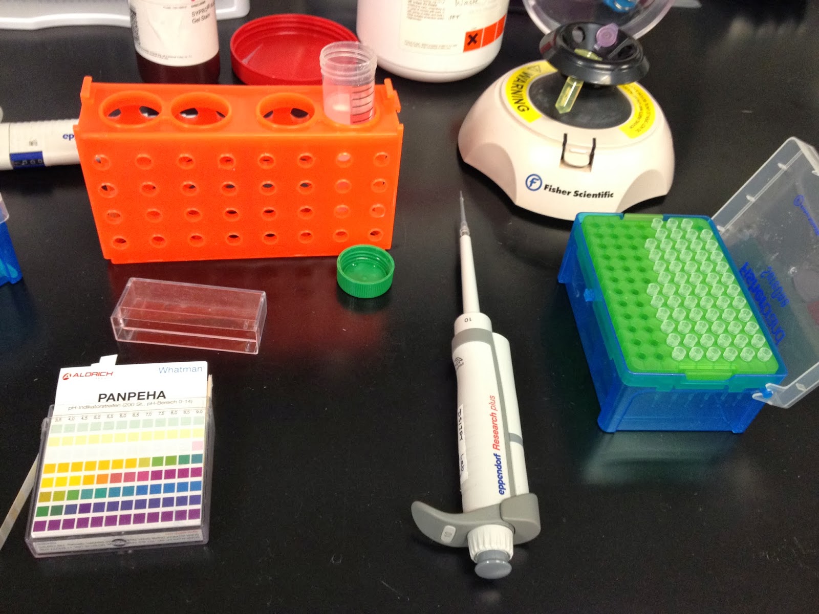
Today, I tested the pH that I could raise the SYPRO dye to before it fell out of the solution. To do this, I tested a 1 mL sample of the dye, adding 1 µL of 2M NaOH at a time and spinning the sample down for 1 minutes to observe if any solid formed.


Once I observed a clear, jellylike solid substance forming from the solution, I used pH strips to test the pH of the solution, which I found to be around 6. I then continued to raise the pH of the dye to pH 10 to see if there were any other effects of increasing the pH. I found that there were no effects other than a clear, jelly-like substance forming at the bottom of the sample. We then wanted to know if any of the dye stayed in the solution. To do this, I took small samples of the original dye, the supernatant that was left after I spun down the sample, and the jelly-like substance. JP and I then observed these samples under UV light to observe the fluorescence of the dye. We found that both the jelly-like substance and the supernatant were about half as fluorescent as the original dye.

I can't wait to continue developing the details of my project next week!

Tuesday, January 21, 2014

Lyophilization

Last Friday (January 17th), I returned to RPI to work more with samples from a peptide synthesis. Today, we went through the process of lyophilization, also known as freeze-drying. The peptide samples were from a synthesis that JP completed earlier, which were already frozen in the freezer. To prepare the peptides for lyophilization, we used liquid nitrogen to freeze the peptides and then covered the tubes with both parafilm and foil. We had to poke holes in the foil and parafilm with a thumbtack for the process to work properly. In the process of lyophilization, we use a machine like the one shown below.


In the process of lyophilization, the peptide tubes are placed in a canister that attaches to one of the spouts of the machine. Inside the center pole of the machine, there is an extremely cold coil that's even colder than the liquid nitrogen. This machine applies both force and extremely cold temperatures to the peptides. As the peptides slowly melt, the pressure causes the water vapor to be transferred to the colder coil, where it condenses. After two or three days, this process eventually removes all of the water from the peptide samples, leaving them freeze-dried with the appearance like a dried sponge or old chewing gum. Lyophilization allows us to turn the peptide samples into a powder, which will be used in future experiments with our synthesized peptides.

Today, we also discussed possible topics for a my own research project. My project will likely have to do with testing blocking buffer solutions. My first task was to check the pH of SYPRO protein gel stain to make sure it's not too acidic to use with peptide microarrays. Using pH strips, I found the pH to be approximately 5.


 I will provide more details of my project in coming weeks. This week, I am tasked with researching details about different blocking buffer solutions that are currently in use. I can't wait to establish the objectives of my project!

Saturday, January 11, 2014

Work-Up

Yesterday, (January 10th), I went to RPI for my first research day of the second semester! I worked on a work up of the peptide synthesis I helped prep for in December. First, I swelled the pellets with ethanol to re-saturate them. Then, I washed each well with TFA:DCM using a step pipette to remove blocking groups. This was the first time I've ever used a step pipette! It dispenses a specified amount of liquid without exposing the inside of the pipette to caustic chemicals such as TFA:DCM.


After TFA:DCM was added to each well, I waited 30 minutes for it to drain, then repeated the TFA:DCM wash process two more times. While I waited for the TFA:DCM to drain, I washed slides in ethanol and made 100mL of 20% ethanol.

After the TFA:DCM drained, I washed the wells with DMF three times to remove the TFA:DCM. I then hooked the well plate up to a vacuum and used the vacuum to drain the plate as I washed the wells with ethanol three more times to remove any remaining TFA:DCM and DMF.

Once the washes were complete, I added ethanol to the bottom of the wells as well as on top, so the peptides are kept in ethanol until they are needed.


I can't wait to return and continue my work!